Posted by Amanda Rickard on May 18, 2017

Reprinted with permission.
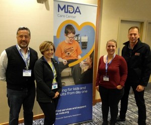
Genea Biocells scientists presented work from four disease modeling projects at the 2017 Muscular Dystrophy Association (MDA) Scientific Conference in Arlington, VA in March. The conference overall was an inspiring and fascinating exhibition of the world’s muscle disease research, with hundreds of attendees working to foster discussion and collaboration toward improving the lives of patients affected by a variety of muscular dystrophies.
Our data on pluripotent stem cell-derived skeletal muscle cells affected by Facioscapulohumeral Muscular Dystrophy (FSHD), a disease which received a lot of attention at the conference. Our work identified three new classes of compounds, all with different molecular targets, that activate DUX4 expression during muscle development. These findings give us interesting insights into the mechanism of DUX4 activation in FSHD, confirm previous findings from other cell systems used for studying FSHD, and reveal druggable targets for DUX4 modulation. Charles Emerson, Jr. (University of Massachusetts) also shared his work, on understanding FSHD muscle development by using pluripotent cells coaxed to become muscle, insights that will help our field to develop more effective therapies and a better understanding of FSHD. Additionally, Peter Jones (University of Nevada) told the audience about FSHD pathophysiology and its role as a model of epigenetic diseases, or diseases that are caused by changes in gene regulation, which we hope will inspire young attendees to join our cause to treat FSHD. Stephen Tapscott (Fred Hutchinson Cancer Research Center) gave those scientists a review of DUX4’s effects on skeletal muscle cells and proposed new mechanisms to stop muscle wasting in FSHD, including interference with DUX4’s toxic effects. Jocelyn Eidahl (Nationwide Children’s Hospital) also taught us more about DUX4 protein chemistry, and how changes to its structure might be harnessed to develop effective therapies. DUX4 can also be targeted in FSHD model mice by RNAi therapy as was presented by Lindsay Wallace (Nationwide Children’s Hospital). Additionally, we learned about the impact and disease burden of FSHD which allows drug developers to target the symptoms that matter most to patients using the most effective clinical trial measures (Jeffrey Statland, University of Kansas Medical Center). Overall, many groups presented their work related to FSHD pathophysiology, model organism development, DUX4 regulation, and therapy development. It’s an exciting time to participate in FSHD research!
We also presented our work in collaboration with Lingjun Rao and Nenad Bursac (Duke University) on using pluripotent stem cell-derived muscle cells to model by Becker and Duchenne Muscular Dystrophies (B/DMD, both caused by mutations in the dystrophin protein). We identified disease markers in 2-D cell models and decreased strength in disease-affected 3-D models by force generation measurement, measures that can be applied to testing therapies. Other MDA attendees taught us plenty about B/DMD with their identification of MRI techniques to identify changes in patients (both Luca Bello, University of Padova and Ami Mankodi, NINDS), efforts to eliminate barriers to genetic testing for patients (Ann Lucas, Parent Project MD), and genes beyond dystrophin that contribute to B/DMD pathogenesis (Elizabeth McNally, Northwestern; Yimin Wang, University of Alabama; Melissa Spencer, UCLA, and others). Much attention was focused on therapeutics testing and development for these diseases, and data from Catabasis Pharmaceuticals, University of Washington, Children’s National Medical Center, WAVE Life Sciences, Sarepta, Boston Children’s Hospital, and others all proposed and tested different approaches.
Research at Genea on pluripotent cell-based models of Myotonic Dystrophy (DM) and Emery-Dreifuss Muscular Dystrophy (EMD) identified disease-specific tissue culture phenotypes that serve as useful measures for cell-based assays and therapeutics testing. Some potential candidates presented at the meeting include inhibition of microsatellite expression to reduce pathogenic RNA in DM (Andrew Berglund, University of Florida) and MBNL1 modulation to rescue DM splicing defects (Fan Zhang, Pfizer). We also learned more about regulation of RNA processing (Eric Wang, University of Florida) and repeat-associated non-ATG translation (Laura Ranum, University of Flordia) in DM cells. Gregory Fedorchak (Cornell) spoke on why and how mutations in EMD cause more fragile muscle nuclei, and Eric Folker (Boston College) presented on nuclear movement and interactions in EMD and healthy muscle cells. Our focus so far at Genea has been EMD caused by LMNA mutations and we were glad to learn more about the effects of EMD-causing emerin mutations in cell-based models as presented by Joseph Ellis and Kimbre Nee (University of the Sciences). We also learned about how mutations in SUN1/2 can cause EMD by impairing nuclear positioning and microtubule connections (Sue Shackleton, University of Leicester), and LMNA mutations in fruit flies cause muscular dystrophy and changes in proteostasis and oxidative stress. All of these contributions lend valuable insights into the pathophysiology of DM and EMD.
Overall the 2017 MDA conference attendees shared many important insights into muscle disease mechanisms and avenues for treatment, leaving us with plenty of work to do when we returned to the lab in San Diego.
For more information on our cell bank feel free to contact us directly at info@geneabiocells.com or visit our website www.geneabiocells.com


Connect with us on social media